
Figure 8. Elevated extracellular Ca2+ inhibits adenosine release during hypoxia and slows the rate of depression of the fEPSP.
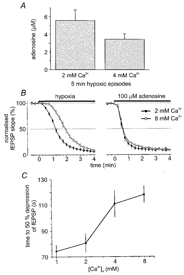
A, sensor measurements of hypoxic adenosine release in 2 mM (n = 16) and 4 mM (n = 4) extracellular Ca2+ showing a trend for reduced adenosine release in high Ca2+. B, in a parallel series of experiments the effect of hypoxia and exogenous adenosine on synaptic transmission in different extracellular Ca2+ concentrations was examined. B left, at time zero hypoxia (black bar) was induced in 2 mM Ca2+ ACSF (filled symbols; n = 18) and 8 mM Ca2+ ACSF (open symbols; n = 7). Note the pronounced retardation in the rate of depression of the fEPSP. B right, in contrast, synaptic transmission was equally sensitive to exogenous adenosine (100 μM; black bar) in both 2 mM Ca2+ ACSF (filled symbols; n = 6) and 8 mM Ca2+ ACSF (open symbols; n = 5). C, concentration dependence of the effects of extracellular Ca2+ on the time to 50 % depression of the fEPSP (1 mM, n = 13; 2 mM, n = 18; 4 mM, n = 14; 8 mM, n = 7). In B and C fEPSPs measured approximately 1 mV.