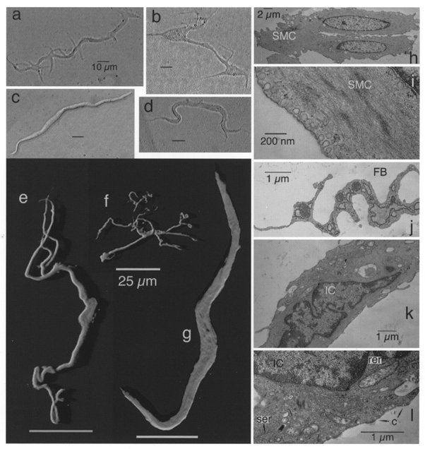
Figure 1. Morphology of cells dispersed from rabbit urethra.

Left panel, interstitial cells (a, b and d) and a typical smooth muscle cell (c) under phase contrast. Shadow projections of confocal stacks of cells which had been incubated with anti-vimentin antibody (e and f) or with anti-myosin antibody (g) are also shown. Right panel, electron micrographs of smooth muscle cells (SMC; h and i), part of a fibroblast (FB; j) and interstitial cells (IC; k and l). (Note caveolae, c, and smooth, ser, and rough endoplasmic reticulum, rer, in l.)
