
Figure 3. All classes of calcium channel couple with equal efficency to exocytosis.
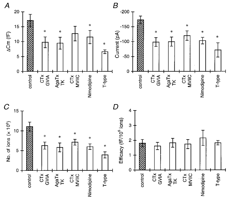
A, the amount of exocytosis per step depolarization for control conditions (total HVA current), selective activation of the T-type current, and the HVA current in the presence of selective HVA Ca2+ channel blockers. First, the mean ΔCm elicited by five step depolarizations was calculated per cell, then the mean for the total group was calculated. Asterisks in A-C indicate statistical differences from the control group ( P < 0.05). B, peak amplitudes of the control Ca2+ current, the T-type current, and the HVA current in the presence of different Ca2+ channel blockers. C, number of Ca2+ ions entering the cell during a step depolarization, calculated as described in Methods. D, efficacy of Ca2+ ions in stimulating exocytosis. ΔCm was divided by the number of Ca2+ ions entering the cell for each depolarizing pulse. For each cell, the mean efficacy for five pulses was calculated. Shown are the means of a number of cells (see text) per group. There was no significant difference between the control group and the groups with the Ca2+ channel blockers. (Stimulation protocols are as described for Fig. 2.)