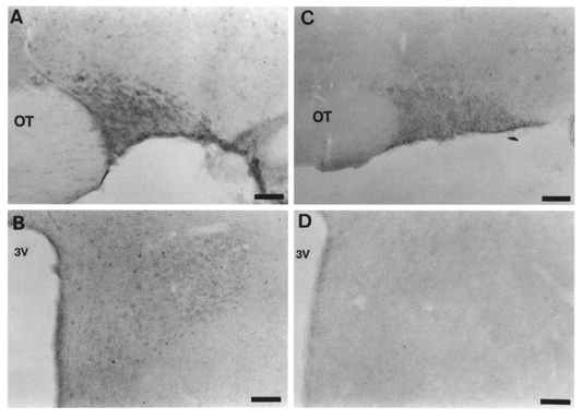
Figure 7. Gzα-like immunoreactivity in the rat SON.

Light micrograph illustrating Gzα-like immunoreactivity in a coronal section of the SON (A) and PVN (B). The staining is found in the perikarya of SON neurones and PVN neurones. This positive staining was abolished by preincubation with Gzα (10−4 M) (C and D). OT, optic tract; 3V, third ventricle. Scale bars, 50 μm.
