
Figure 1. Phase shift of the human circadian pacemaker and acute suppression of plasma melatonin.
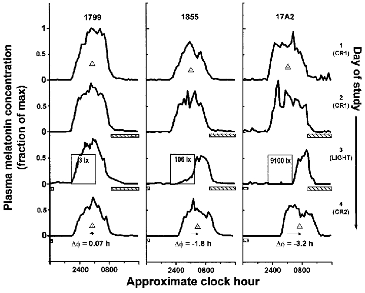
Melatonin profiles during days on which the first constant routine (CR) (1 and 2), the single experimental light exposure 6.5 h in duration (3), and the second CR (4) occurred are shown for three representative subjects (1799, 1855, 17A2). In the dimmest light condition, exposure to the dim light stimulus (∼3 lx) had little effect on either the phase of the melatonin rhythm (phase shift (ΔΦ) 0.07 h) or concentration of plasma melatonin (suppression 11 %). In the brightest light condition (∼9100 lx), light both shifted the rhythm (ΔΦ -3.2 h) and completely suppressed plasma melatonin (98 %). Exposure to dim room light (∼106 lx) evoked more than half of the shift observed in the brightest light condition (ΔΦ -1.8 h compared with -3.2 h) and a nearly equal amount of suppression (88 %). During the CRs and day of experimental light exposure, subjects were exposed to no more than 5 lx in the horizontal angle of gaze at any time except during the scheduled sleep episodes (hatched bars < 0.03 lx) and the experimental light exposure (labelled open boxes, see Fig. 2). Individual subject data were plotted on a time scale in which their habitual wake time was assigned a reference value of 08.00 h. Phase of the melatonin maximum (midpoint of the upward and downward mean crossings) during each CR is noted as the ▵. For graphical purposes, ordinate values were normalized to each subject's absolute peak plasma melatonin concentration.