
Figure 3. Response of a CMH and a CMiHi fibre to repetitive electrical stimulation with interpolated conditioning stimuli at different interstimulus intervals.
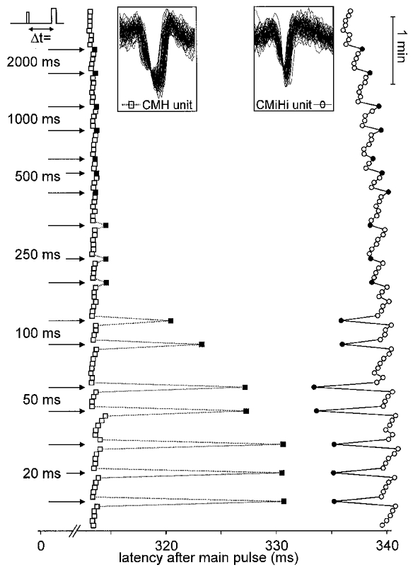
Only action potentials (APs) are displayed, from top to bottom in successive order of traces symbolised by squares (CMH fibre) or circles (CMiHi fibre) The superposition of all APs in the insets shows their stable shape. Segments 313–342 ms after each stimulus are shown. Intracutaneous electrical stimuli were applied at 4 s intervals (0.25 Hz). Arrows on the left indicate additional pulse interposition at the given ISIs (Δt). Filled symbols represent the first AP after the additional pulse.