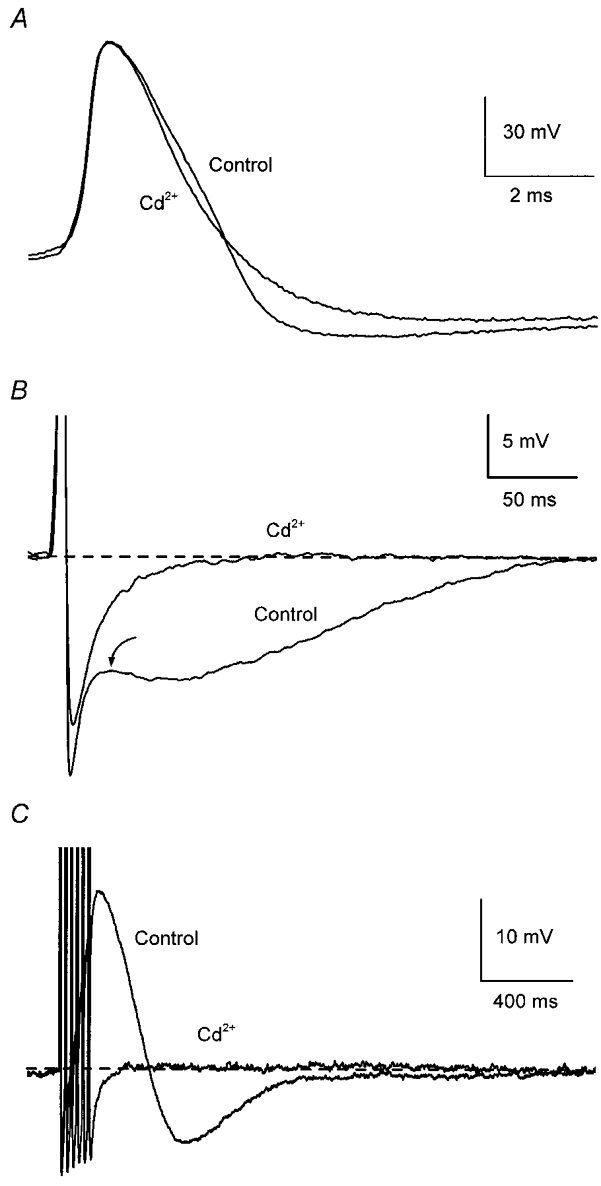
Figure 1. Ca2+-dependent afterpotentials in mouse sympathetic ganglion cells.

Records from a neurone in the superior cervical ganglion. A, addition of 500 μm Cd2+ reduced action potential (AP) half-width without affecting its amplitude. B, addition of Cd2+ abolished the depolarizing inflection following AP repolarization (arrow) and markedly reduced the afterhyperpolarization (AHP) following a single AP. C, addition of Cd2+ abolished the afterdepolarization (ADP) and reduced the AHP after a train of six APs at 40 Hz. APs in B and C are truncated (as in subsequent figures) and traces in control and test solutions have been superimposed with labels placed closest to the relevant trace. In these, and in the current-clamp records in subsequent figures, the dashed line indicates the baseline membrane potential, which was maintained close to −55 mV by passing current through the microelectrode.
