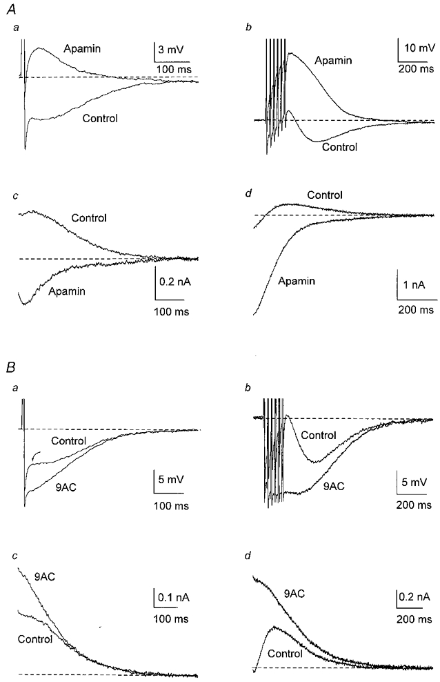
Figure 2. Apamin and 9AC reveal the Cl− and K+ currents underlying afterpotentials.

Responses following a single AP are shown on the left (a) and after a train of six APs at 40 Hz on the right (b) in this and subsequent figures. A, in one cell, addition of apamin (100 nm in this and subsequent figures) abolished the slow AHP, revealing an ADP after one AP (a) and enhancing the ADP after a train of six APs at 40 Hz (b). In the same neurone, outward tail currents following a single AP (c) or a train of APs (d) were blocked by apamin, revealing inward tail currents. B, in another cell, addition of 9AC (2 mm in this and subsequent figures) reduced the depolarizing inflexion in the AHP after a single AP (a) and the small ADP following a train of APs (b), enhancing the AHP. Arrow points to the transition from fast to slow AHP. In the same neurone, 9AC blocked the inward currents after a single AP (c) and after a train of APs (d), enhancing the outward tail currents. In these, and in voltage-clamp records in the following figures, the dashed line indicates the holding current needed to keep the membrane potential at −55 mV. In this, and in the following figures except Fig. 5Ab, the trains consisted of six APs at 40 Hz.
