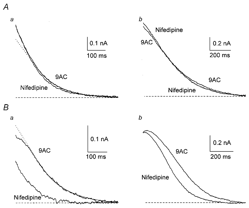
Figure 6. L-type Ca2+ channels contribute to activation of gK,Ca1 in a subgroup of neurones.

Two groups of cells were identified on the basis of the time course of the outward current recorded in the presence of 9AC. A, response of type I cells. When an exponential function was fitted to the outward current, starting 150 ms after the AP (continuous line), and extrapolated to the initial part (dotted line in this and subsequent figures), the outward current of type I cells either fitted the exponential well or initially fell above it (a). When 10 μm nifedipine was added, the outward current from a type I cell was not affected after one AP (a) and showed a small increase in peak current after a train (b). B, response of type II cells. The outward current of type II cells initially fell below the extrapolated exponential (a). In one type II cell, nifedipine markedly reduced gK,Ca1 after one AP (a) and had a smaller effect on the latter part of the current after a train (b).
