
Figure 4. Immunohistochemical evidence for the presence of dKv3.1 in dog atrial myocytes.
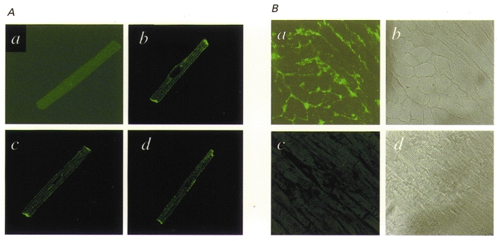
A, immunocytochemical images of canine atrial myocytes prepared with primary antibody pre-incubated (a) and not pre-incubated (b-d) with the Kv3.1 peptide against which the antibody had been raised. B, tissue sections of dog atrium: a is a typical immunohistochemical image prepared with Kv3.1 antibody and b is a bright-field micrograph of the same section; c was prepared in the same fashion as a, but with the Kv3.1 antibody pre-incubated with Kv3.1 peptide, and d is a bright-field image of the section shown in c.