
Figure 6. Voltage- and time-dependent properties of dKv3.1 currents in oocytes.
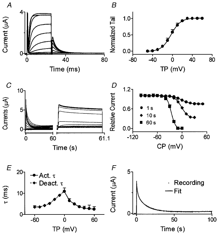
A, currents elicited by 20 ms pulses from a Vh of −80 mV. Tail currents were recorded upon repolarization to −30 mV. B, normalized tail currents fitted by a Boltzmann distribution (n = 8). C, currents elicited by 60 s conditioning pulses to conditioning potentials (CP) between −100 and +50 mV followed by a 1100 ms test pulse to +50 mV. D, inactivation curve with conditioning pulses of 1, 10 and 60 s (n = 8 for each). E, voltage-dependent activation and deactivation time constants (n = 8). F, time-dependent inactivation of dKv3.1 during a 100 s pulse to +40 mV and biexponential fit.