
Figure 8. Evaluation of dKv3.1 protein and IKur,d current expression in dog ventricle.
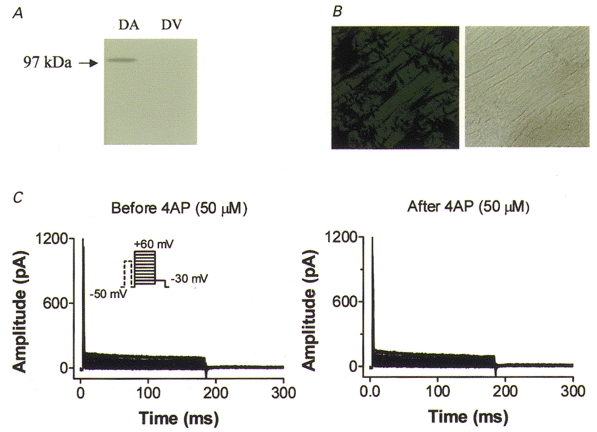
A, Western blots of dog atrial (DA) and ventricular (DV) tissues with Kv3.1 antibody. B, immunohistochemical image obtained with Kv3.1 antibody (left) and bright-field image (right) on a dog ventricular tissue section. C, currents recorded from a dog ventricular myocyte with the same voltage protocol and conditions as used to record IKur,d in atrium (Fig. 5B). Currents were recorded before (left) and after (right) superfusion with 50 μm 4-AP.