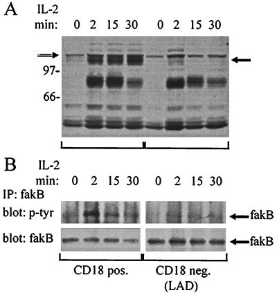
Figure 4.
IL-2 induces tyrosine phosphorylation of fakB in CD18-positive but not in CD18-negative T cells. (A) A CD18-positive T cell line from a healthy donor (left four lanes) and a CD18-negative T cell line from a LAD patient (right four lanes) were incubated with medium or IL-2 (15 ng/ml) for the periods of time indicated. Cells were then lysed and IL-2-induced tyrosine phosphorylation was analyzed by Western blotting with an anti-phosphotyrosine mAb (4G10). The positions of prestained molecular standards (Amersham) are indicated on the left in kDa. A solid arrow (←) indicates the IL-2-inducible 125-kDa phosphotyrosine protein (p125) likely to represent fakB (compare below), whereas an open arrow (⇒) indicates a constitutively tyrosine phosphorylated 130-kDa protein likely to represent pp125FAK. (B) CD18-positive (Left) and -negative (Right) T cell lines were stimulated with IL-2 as in A. Lysates were immunoprecipitated with anti-fakB antibody and immunoblotted with anti-phosphotyrosine mAb (Upper) or anti-fakB antibody (Lower). These data are representative of three independent experiments.

