
Figure 5. Inhibition by quinpirole of α1B currents, in the presence or absence of co-expressed VDCC β subunits.
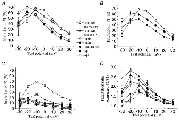
A, the percentage inhibition by quinpirole in P1 for currents formed by α1B/α2δ without co-expressed VDCC β subunits, or with the VDCC β1b, β3 or β4 subunits, as shown in the key. Inhibition was measured over the −30 to +30 mV range. The control values for the 50 and 200 ms prepulse (PP) data were not significantly different and have been combined (○). B, the percentage inhibition by quinpirole in P1 for currents formed by α1B/α2δ without co-expressed VDCC β subunits, or with the VDCC β2a, or C3,4S-β2a subunits, symbols as shown in the key. The controls for the 50 and 200 ms prepulse (PP) data were not significantly different and have been combined (○). C, the percentage inhibition in P2, after a depolarizing prepulse of 50 or 200 ms duration, as stated, for the same combinations of subunits as in A and B. D, facilitation ratio (as corrected P2/P1, see Methods) for the different subunit combinations (see key), between −30 and +30 mV. The numbers of experiments are as follows for both A and B:α1B α2δ (50 ms prepulse), 9; α1B α2δ (200 ms prepulse), 4; +β1b, 11; +β2a, 20; + C3,4S-β2a, 8; +β3, 12; +β4, 12.