
Figure 1.
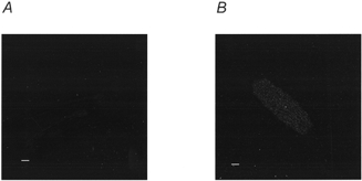
Confocal image of cells permeabilised in the presence and absence of fluorescent S2
Sections were taken through the mid-line plane of representative rat ventricular myocytes following streptolysin O permeabilisation in the absence (A) and in the presence (B) of tetramethylrhodamine isothiocyanate-labelled S2. Permeabilisation and recording of images were performed in parallel. Scale bars represent 10 μm.