
Figure 3.
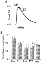
The effect of permeabilisation in the presence of the S2 fragment of myosin on [Ca2+]i transients in fura-2-loaded rat ventricular myocytes
A, fast time base recordings of representative [Ca2+]i transients obtained in control permeabilised (CP) and S2 permeabilised (S2) myocytes. [Ca2+]i is expressed as fura-2 fluorescence ratio units (RU). B, mean data showing diastolic [Ca2+]i (Dias), systolic [Ca2+]i (Sys), amplitude of the [Ca2+]i transient (Amp), time to peak of the transient (TTP) and time to half-recovery of the transient (t1/2) in control cells (□; n = 31), streptolysin O-permeabilised cells ( ; n = 39) and cells permeabilised in the presence of S2 (
 ;n = 39). All values are expressed as means +s.e.m. as a percentage of that recorded in control cells. Control values for the parameters were: diastolic [Ca2+]i, 0.111 ± 0.004 RU; systolic [Ca2+]i, 0.216 ± 0.012 RU; [Ca2+]i transient amplitude, 0.105 ± 0.009 RU; time to peak, 94 ± 6 ms; time to half-recovery, 328 ± 18 ms. †P < 0.05 compared with control (unpermeabilised) cells; *P < 0.05 compared with control permeabilised cells (Student's t test with Bonferroni correction).