
Figure 6.
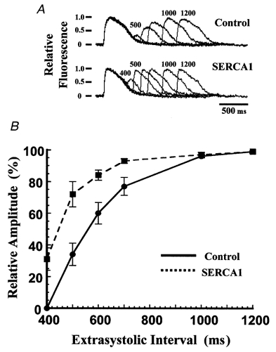
Time course of refilling of Ca2+ stores in embryonic chicken cardiac myocytes
A, chicken cardiac myocytes loaded with fluo-4 were stimulated with a two-pulse protocol after pacing at 1·0 Hz. The last paced transient and the extrasystolic transient were recorded while varying the extrasystolic interval. Each record was normalized to its conditioning transient and superimposed as a series. Upper series, control myocyte. Lower series, myocyte overexpressing SERCA1. Time intervals are indicated above the traces (in ms). B, relative amplitudes of the Ca2+ transients as a function of the extrasystolic interval. Amplitude was calculated as the difference between the peak of the transient and the baseline fluorescence just prior to the stimulus pulse. Points are means ± s.e.m. •, control cells (n = 20); ▪, cells overexpressing SERCA1 (n = 16).