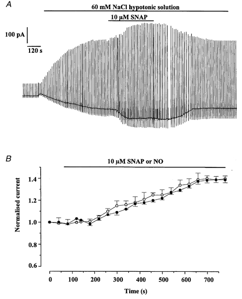
Figure 3.

Cells in which SNAP and NO increased Iswell
A, typical cell showing the increase of Iswell by 10 μm SNAP. B, time dependence of the potentiating effect of SNAP (○) and NO (•) on Iswell recorded at −50 mV. Each point is the mean ±s.e.m. of 9 cells for SNAP and 6 cells for NO.
