
Figure 1.
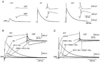
DRP and CDP sensitivity to stimulus intensity and GABAA, AMPA and NMDA receptor antagonists
A, DRP evoked by dorsal root stimulation at 1.5, 2 and 50 times threshold (T). Insets show simultaneously recorded CDP and DRP at a faster sweep speed. Arrows indicate the time of stimulus application B, DRP at 50T in normal medium (control) and after sequential application of 40 μm bicuculline (Bic), 100 μm AP-5 (AP-5 + Bic) and 25 μm CNQX (CNQX + AP-5 + Bic). The simultaneously recorded CDP is shown in the insets at a faster sweep speed. C, DRP at 50T, in normal medium (control) and after sequential addition of 25 μm CNQX and 10 μm strychnine (CNQX + Stry), 40 μm bicuculline (Bic + CNQX + Stry), and finally 100 μm AP-5 (AP-5 + Bic + CNQX + Stry). Inset, simultaneously recorded CDP at a faster sweep speed. In this and subsequent figures, negative polarity is up and the DRP was recorded in DC mode. A and C are from the same preparation.