
Figure 5. Inward currents in cultured HISM cells developing in divalent cation-free external solution.
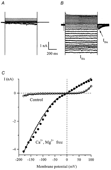
A and B, superimposed current traces elicited by 800 ms duration voltage steps from −40 mV to test potentials between +100 and −180 mV in 10 mV increments in control (130 mm Na+, 2.5 mm Ca2+, 1.2 mm Mg2+; A) and after external Ca2+ and Mg2+ removal (B, denoted as IHA). Note the instantaneous activation and deactivation of IHA as well as the noisy appearance of the current at negative potentials. The current seen upon repolarization to −40 mV is denoted as IDA. C, I-V relationships for currents measured at the end of the pulse (○ and •) and, in the same cell, by applying 1 s duration voltage ramps from +100 to −180 mV (continuous lines). The ramp protocol is illustrated in Fig. 6A.