
Figure 7. Effects of carbachol on IHA and IDA.
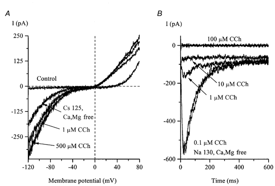
A, the control I-V relationship was measured as described for Fig. 6C. After IHA had developed in divalent cation-free, high-Cs+ external solution, it was rapidly potentiated by carbachol (CCh) application. IDA was abolished in Cs+-containing external solution and was inactivated at −40 mV. B, in a different cell, IDA measured at −40 mV in divalent cation-free, high-Na+ external solution was strongly inhibited by carbachol.