
Figure 8. Pharmacological properties of IHA and IDA.
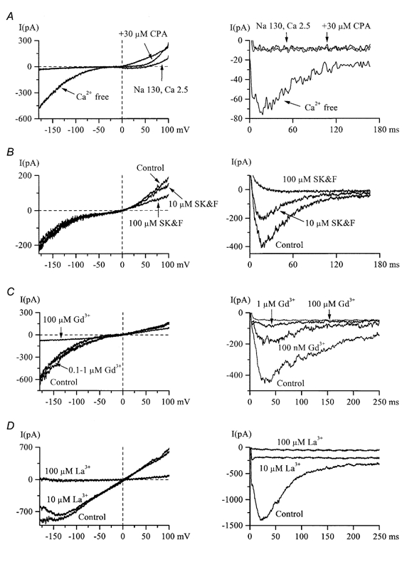
A, 5 min treatment with 30 μm CPA failed to induce an inward IHA (left) or IDA (right) in 2.5 mm Ca2+, high-Na+ external solution (n = 5), but both currents were readily activated following Ca2+ removal. Note that at positive potentials current could be activated by CPA. B, SK&F 96365 at concentrations of up to 100 μm did not inhibit inward IHA (left), but completely abolished IDA (right) in the same cell (n = 7). Current at positive potentials at which it could be activated by CPA (left panel in A) was also inhibited by SK&F 96365. C and D, IDA (right) was more sensitive to the inhibitory action of Gd3+ or La3+ compared to IHA (left) but both currents were abolished by 100 μm of either blocker. In all panels IHA and IDA were measured in the same cells.