
Figure 8. Presence of KATP channels in δ-cells.
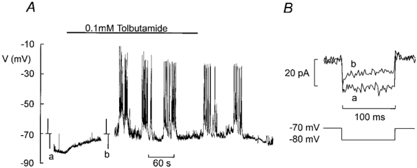
A, electrical activity recorded from a δ-cell (identified by the presence of Na+ current and absence of an A-current) in the absence of glucose before and after addition of 0·1 mm tolbutamide (indicated by horizontal line). At the time points indicated by a and b, the recording was interrupted, the amplifier switched into voltage-clamp mode and ±10 mV pulses were applied from −70 mV to monitor membrane conductance. B, membrane currents evoked by 10 mV hyperpolarising pulses in the absence (a) and presence (b) of 0·1 mm tolbutamide. The current amplitude was measured after the capacitive transients had decayed.