
Figure 3. Behaviour of the two γs motor axons from Fig. 1 during rhythmic stepping movements.
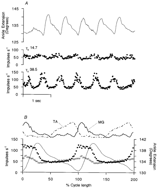
In A the instantaneous γ-axon firing frequencies and ankle rotation are shown in real time (same γs axons as in Fig. 1). The γs motor axon conduction velocities are shown in m s−1. The upper trace is ankle rotation with extension (plantar flexion) upwards. The angle is measured between the front of the tibia and the dorsum of the foot. In B, cycle averages are computed for 14 cycles of regular amplitude of ankle movement from the data illustrated in A. The two top traces are the smoothed, rectified EMG signals from MG and TA. The lower three records are the averaged frequency records for a type-1 γs axon (•) and a type-2 γs axon (○) and the averaged ankle angle (continuous line). In the horizontal scale, time is normalised with respect to the individual stepping cycle durations. The mean cycle period was 0.65 s.