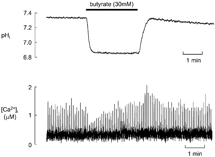
Figure 1. The effect of butyrate (30 mM) on pHi (top) and [Ca2+]i (bottom) in representative rat ventricular myocytes.

The cells were field stimulated at 0.2 Hz and exposed to butyrate for 2 min as shown above the traces. Different cells were used for the pH and [Ca2+]i measurements. In this (and all other) figures dimethylamiloride (20 μM) was present throughout to inhibit Na+-H+ exchange.
