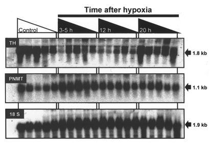
Figure 3. Autoradiographs of Northern blots of total adrenal RNA from fetal sheep exposed to acute hypoxia.

Representative autoradiographs of a Northern blot after hybridisation of radiolabelled TH cDNA, PNMT and 18 S antisense oligonucleotide probes with total RNA (20 μg lane−1) extracted from adrenal glands collected from fetal sheep at 129-144 days gestation after experimental normoxia (Control, indicated by area under open triangle, n = 5) or 3-5 h (n = 5), 12 h (n = 5), and 20 h (n = 5) after 30 min of experimental hypoxia (indicated by the areas under the filled triangles). The approximate sizes of the relevant transcripts are indicated on the right-hand side of the panels.
