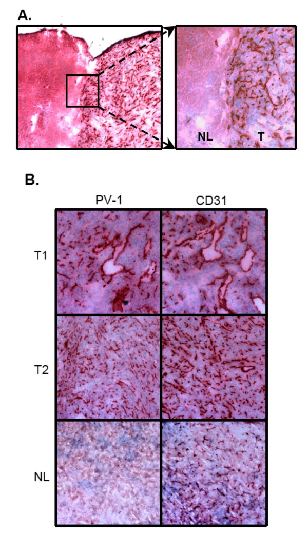
Figure 2.
PV-1 protein expression in intracranial glioma xenografts. A. Immunohistochemistry for PV-1 was performed on frozen brain sections containing U87:U87/VEGF tumors (T1, T2) or normal brain (NL); 100×. PV-1 expression was detected solely on the microvessels within the tumors and showed a comparable staining pattern to that of the endothelial control marker, CD31. PV-1 expression was not detected in normal brain as compared to CD31. B. The interface between normal brain (NL) and brain tumor (T) demonstrated that PV-1 expression was tightly restricted to the tumor; overview 40×, detail 100×.

