Fig. 1.
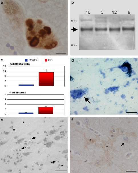
Neuronal pentraxin II (NPTX2) is strongly upregulated in substantia nigra and cerebral cortex of PD patients. a NPTX2 immunocytochemistry demonstrating specific staining of Lewy bodies in a dopaminergic neuron. b Western blotting confirms specificity of the anti-NPTX2 antibody. c qRT-PCR shows highly increased levels of NPTX2 mRNA in substantia nigra and, to a lesser extent, in frontal cortex. The arbitrary units along the ordinate represent relative fold changes [25]. The control value is 1; error bars indicate SEM. d, e In situ hybridisation demonstrates NPTX2 mRNA expression in nerve cells as well as glia in both substantia nigra and frontal cortex. Labelling is also found over non-pigmented neurons (arrow in d, SN). The arrows in e mark glial cells (cortex). In addition, there is autoradiographic signal in the neuropil (asterisks) which would be in keeping with the presumed dendritic expression of NPTX2 mRNA. f NPTX2 immunocytochemistry of cortical neuropil reveals occasional intercellular patches of “arborised” labelling also possibly suggestive of a dendritic localisation (asterisks). The arrow points to a NPTX2 immunoreactive glial cell. Counterstaining in a and f, haemalum, and toluidine blue in d. Scale bars: 20 μm (a, f), 50 μm (d), and 100 μm (e)

