Fig. 2.
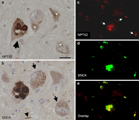
Co-localisation of NPTX2 and alpha-synuclein. a, b NPTX2 and alpha-synuclein immunolabelling of adjacent tissue sections demonstrates significant but not complete overlap between the two proteins. The large arrow in a denotes the same Lewy body-containing dopaminergic nigral neuron. Small granular cytoplasmic deposits showing alpha-synuclein immunoreactivity in some nerve cells (arrows in b) are NPTX2 negative. These aggresomes are sometimes difficult to distinguish from neuromelanin granules based on staining alone but their cytoplasmic distribution is also different from the latter. In contrast, Lewy neurites are double-labelled (arrow head in b). Neuromelanin (asterisks in a) is unstained. c, d, e Dual immunofluorecence demonstrates co-localisation of neuronal pentraxin II (red, c) and alpha-synuclein (green, d). Double-labelled structures (arrows in c) light up in yellow in the overlay (e). Scale bar: 40 μm (a, b), and 80 μm (c–e), respectively

