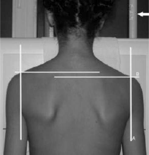
Fig. 2.
Clinical picture used to evaluate clinical shoulder balance. aVertical lines were drawn through the posterior axillary folds. b The height difference between the horizontal lines where vertical lines intersected with the shoulders was measured to reflect the clinical shoulder balance (arrow the tube whose size was used to calibrate the clinical shoulder balance measurements)

