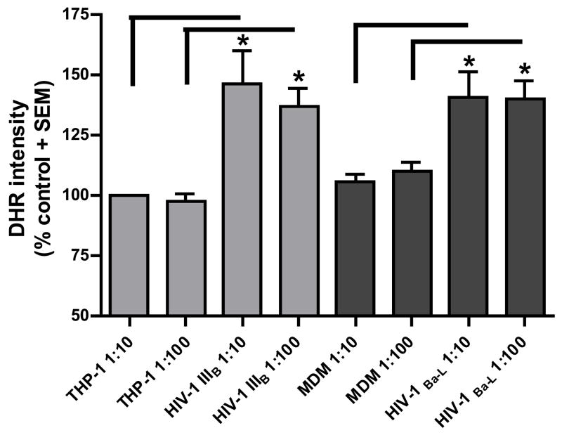
Figure 3. Effect of supernatants from HIV infected monocytic cells on mitochondrial function of DRG cell bodies.
Mitochondrial free radical production and membrane potential was measured in human DRG cell bodies following treatment with supernatants from THP-1 cells infected with HIV-1IIIB and MDM infected with HIV-1Ba-L.
A) Increase in DHR intensity is shown as standardized data. * Supernatant from infected monocytic cells versus the matched control, p < 0.05. Each data bar represents approximately 100 analyzed neurons
B) Mitochondrial membrane potential (red/green ratio) is shown as standardized data. * Supernatant from infected monocytic cells versus the matched control, p < 0.05. Each data bar represents at least 150 analyzed neurons.


