
Figure 6. Detection of 3NT modified proteins in peripheral nerve of HIV infected patients.
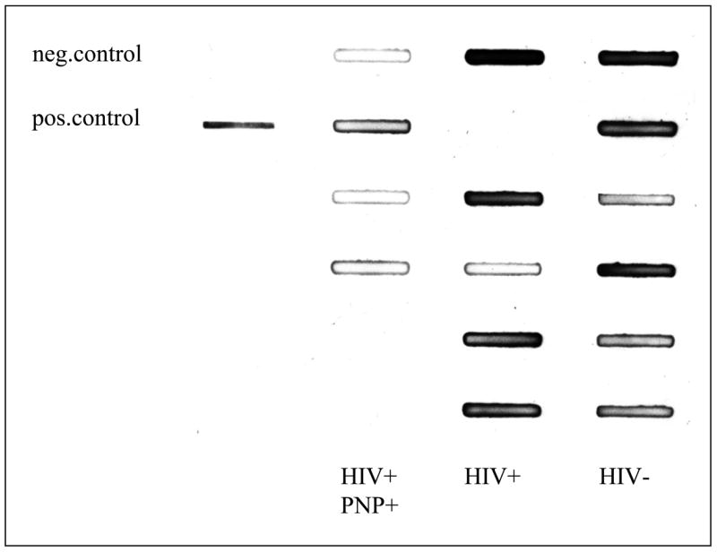

A) Sciatic nerve extracts were analyzed by slot blot by using an antisera to 3-NT. The left row represents a negative (BSA) and a positive control (nitrosylated BSA), the second row shows 5 sciatic homogenate samples from HIV-seropositive individuals with neuropathy. The slots in the third row were loaded with samples from HIV-seropositive subjects without neuropathy and the right row with samples from the HIV-seronegative controls
B) The bar graph represents the mean area±SEM. ** p< 0.05 compared to the HIV-negative controls.