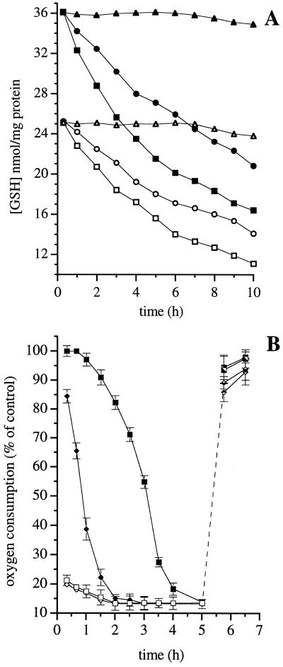
Figure 4.
Mechanism of inhibition of respiration at complex I by DETA-NO. (A) Changes in GSH concentration were measured in control cells (triangles) or in cells treated with DETA-NO (0.1 or 0.5 mM, circles and squares, respectively) after an 18-h incubation in culture medium in the absence (filled symbols) or presence (open symbols) of l-BSO (0.3 mM). Results shown are representative of two experiments. (B) J774 cells were incubated for 18 h in the culture medium in the absence (squares) or presence of l-BSO (0.3 mM, diamonds) and then treated with DETA-NO (0.5 mM). Oxygen consumption was measured before (open symbols) or after (filled symbols) addition of Hb (8 μM). After 5 h of incubation, DETA-NO was removed by washing and the cells were incubated with GSH–methylester (1 mM; filled symbols, Lower) or exposed to a high intensity light (filled symbols, Upper; n = 3).

