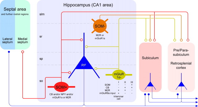
Figure 7.
Diagram showing the novel subsets of CA1 hippocampal GABAergic neurons projecting to the septal and/or retrohippocampal areas. The in vivo single-cell labeling and retrograde tracer experiments suggest three main types of hippocampal GABAergic projection neuron. The first major population (red) is located in str. oriens, projects to both the retrohippocampal and septal areas, and is SOM and mGluR1α positive. The second population (green) is less common in str. oriens, projects exclusively to the subicular areas, and shows diverse molecular expression profiles, as indicated below the cell. Some cells of this group receive strongly mGluR8a-positive boutons and constitute the trilaminar cell type. The two subsets of projection neurons in str. oriens fire at or shortly after the trough of theta oscillations and increase their firing rate during sharp wave-associated ripple oscillations. Their local targets in the CA1 area are mainly small-diameter dendrites of pyramidal cells, but up to a quarter of the postsynaptic elements of an individual neuron can be other interneurons. The third population (brown), found in the str. radiatum and lacunosum-moleculare, projects to retrohippocampal areas but not to the septum; these neurons are SOM negative but often positive for M2R or mGluR1α. They fire at the descending phase of theta oscillations but are not activated during sharp wave-associated ripples. Note that the subiculum receives both glutamatergic pyramidal cell and GABAergic nonpyramidal cell input, but other areas may receive only GABAergic input from the CA1 area. GABAergic terminals are shown as open circles, and glutamatergic terminals are shown as filled circles. Only the major molecular markers and most frequent cells are included for simplicity. slm, Str. lacunosum-moleculare; so, str. oriens; sp, str. pyramidale; sr, str. radiatum

