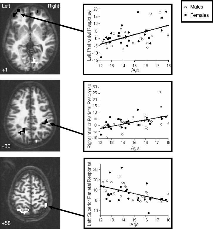
Fig. 2.
Brain regions showing significant relationships between age and fMRI response to spatial working memory relative to vigilance across adolescence. Black clusters indicate areas showing a positive relationship between age and fMRI response, and white regions represent clusters showing a negative relationship between age and fMRI response (p < .05, cluster volume > 943 microliters). Numbers below images refer to axial slice positions.

