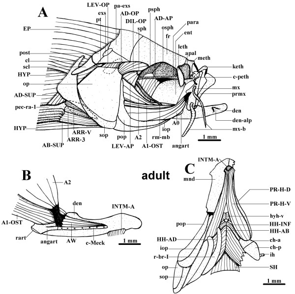
Figure 5.
Adult cranial musculature. A. Lateral view of the cranial cephalic muscles and surrounding skeletal structures of an adult zebrafish (45.1 mm TL). B. Mesial view of the left mandible and adductor mandibulae of an adult zebrafish (45.1 mm TL), part of the anterior intermandibularis is also shown, the adductor mandibulae A0 was removed. C. Ventral view of the cephalic muscles and surrounding skeletal structures of an adult zebrafish (45.1 mm TL), on the right side a portion of the hyohyoidei adductores, as well as of the mandible, was cut, and the opercle, interopercle, subopercle and preopercle are not represented. A0, A1-OST, A2, AW, sections A0, A1-OST, A2 and Aω of the adductor mandibulae; AB-SUP, abductor superficialis; AD-AP, adductor arcus palatini; AD-OP, adductor operculi; AD-SUP, adductor superficialis; angart, angulo-articular; apal, autopalatine; ARR-3, arrector 3; ARR-V, arrector ventralis; c-Meck, Meckelian cartilage; c-peth, pre-ethmoid cartilage; ch-a, ch-p, anterior and posterior ceratohyals; cl, cleithrum; den, dentary bone; den-alp, anterolateral process of dentary bone; DIL-OP, dilatator operculi; ent, entopterygoid; EP, epaxialis; exs, extrascapular; fr, frontal; HH-AB, hyohyoideus abductor; HH-AD, hyohyoidei adductores; HH-INF, hyohyoideus inferior; hyh-v, ventral hypohyal; HYP, hypaxialis; ih, interhyal; INTM-A, intermandibularis anterior; iop, interopercle; keth, kinethmoid; leth, lateral-ethmoid; LEV-AP, levator arcus palatini; LEV-OP, levator operculi; meth, mesethmoid; mnd, mandible; mx, maxilla; mx-b, maxillary barbel; op, opercle; osph, orbitosphenoid; pa-exs, parieto-extrascapular; para, parasphenoid; pec-ra-1, pectoral ray 1; pop, preopercle; post, posttemporal; prmx, premaxilla; PR-H-D, PR-H-V, dorsal and ventral sections of protractor hyoidei; psph, pterosphenoid; pt, pterotic; r-br-I, branchiostegal ray I; rart, retroarticular; rm-mb, mesial branch of ramus mandibularis; scl, supracleithrum; SH, sternohyoideus; sop, subopercle; sph, sphenotic.

