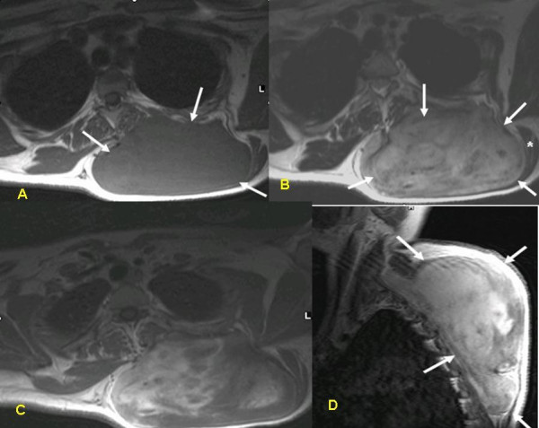
Figure 2.

MRI of the tumor: T1W pre-(A) and post-(B) gadolinium injection, T2W (C) and T1W post gadolinium, sagittal view (D). The tumor (arrows) has a heterogenous appearance on T2W images and enhances with the injection of contrast material, demonstrating its vascularity. It is located beneath the trapezius muscle (asterisk) which is atrophic. The paraspinal muscle is compressed medially.
