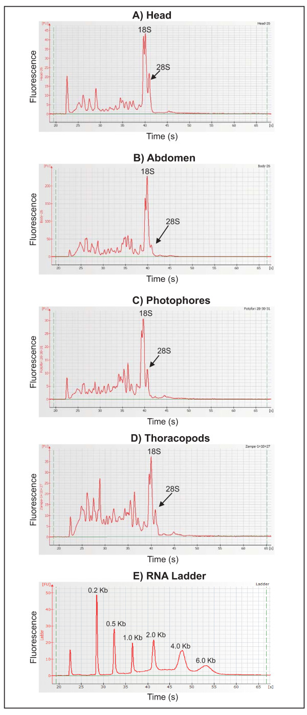
Figure 1.
Electropherograms of E. superba tissue-specific total RNAs. (A-D) Electropherograms resulting from Agilent 2100 bioanalyzer analysis on total RNA extracted from head, abdomen, photophores and thoracopods. X-axis: time of ribosomal RNA peak appearance, corresponding to the size of the fragment; Y-axis: fluorescence of the peak, corresponding to its concentration. The size and the concentration of the sample peaks are calculated by the software via comparison with a RNA ladder at known concentration (E). E. superba RNA samples showed some products with a migration time between 22 and 35 seconds (from 200 bp to 1.000 bp) indicating a partial RNA degradation.

