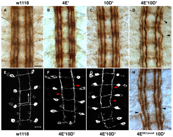
Figure 3.
CNS phenotype of Ptp4E Ptp10D double mutants. (a-d,h) Stage 17 embryos were stained with mAb 1D4 and visualized using HRP immunohistochemistry. (e-g) Stage 17 embryos expressing the transgene SemaIIb-τmyc were stained with anti-Myc antibodies, and images are projections of confocal stacks. (a) Wild-type embryos at stage 17 show three distinct longitudinal tracts on either side of the midline. Commissural bundles do not stain with 1D4 at this stage, although cell bodies showing light staining are visible here. Ptp4E1 (b) and Ptp10D1 (c) embryos do not show any defects in CNS axon guidance, and are indistinguishable from control w1118 CNS (a). The double mutants Ptp4E1 Ptp10D1 (d) and Df(1)ovo4 ptp10D1 (h) show mild CNS defects. The longitudinal axons appear wavy and the outermost longitudinal axon tract is discontinuous and often invades the middle (intermediate) longitudinal axon tract (arrowheads). SemaIIB axons were visualized using the SemaIIb-τmyc transgene. (e) In the wild-type CNS, semaIIb-τmyc expressing axons project across the midline along the anterior commissural bundle and extend anteriorly along the intermediate 1D4 fascicle (arrows indicate the direction of axon trajectory). (f,g) SemaIIb-τmyc axons project in a normal manner in the double mutants. However, they have a consistent defect within their longitudinally projecting segment, in which the axon bundles do not form a tight bundle but have a frayed appearance (arrowheads). Anterior is up in all panels. Scale bars are 10 microns.

