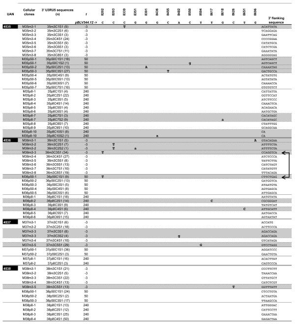
Figure 3.
Somatic (miniscule letters) versus reverse-transcriptase (capital letters) associated substitutions of the BLV 3' RU5 sequence during early experimental sheep infection. Overall, 65 distinct BLV 3' integration sites were isolated; the first 8 bases of the corresponding flanking cellular sequences are given on the right. RU5 sequences were aligned according to the sequence of the wild type BLV sequence 344 used for experimental infection of animals 4535 and 4536. Sheep are identified by their unique animal number (UAN). Each cluster of RU5 sequences sharing a common integration site, and therefore belonging to a unique clone of expanded B cells, is identified by its cellular clone number. For each cellular clone the number of non-unique 3' U3RU5 consensus sequences is indicated between brackets in the third column. Cellular clones harboring a mutated 3'U3RU5 sequence are overlined in grey. A horizontal double bar separates the clusters of sequences derived from each of the 4 sheep DNA samples. For each animal, cellular clones are sorted according to their date of isolation, i.e. 3 days before, 50 after seroconversion and 240 days after experimental infection. The two horizontal arrows represent the two times at which the same C8202T substitution was observed in 2 distinct sequences deriving from animal #4536.

