To the Editor,
Case study: An 88-year-old, black man was referred to a gastroenterologist for evaluation of massive ascites causing severe abdominal distension, nausea, vomiting, bloating, and weight gain (26 lb in 9 months). Past medical history was significant for prostate cancer status postsurgical resection (1988), hyperlipidemia, degenerative joint disease, hypertension, chronic renal insufficiency, normal colonoscopy evaluation (2005), and laparotomy (April 2006) for resection of a midabdominal mesenteric mass that was diagnosed as sclerosing mesenteritis on histopathology. The patient was well until 6 months later, when he developed extensive ascites. The patient was referred to a gastroenterologist for the evaluation of new-onset progressively worsening ascites causing abdominal discomfort and distension. At the time of presentation, the patient was taking pravastatin, furosemide, cimetidine, potassium, colchicine, and prednisone. The patient had no known allergies to any medication or substance. He was a married, retired letter carrier living at home with his wife and had quit smoking 37 years ago. There was no significant family history. Review of systems was notable for nausea, occasional vomiting, and difficulty ambulating even 1 flight of steps.
On examination, the patient appeared ill with no acute distress with stable vital signs. The remainder of the exam was significant for decreased bilateral basal breath sounds, a well-healed median laparotomy scar with a markedly distended abdomen notable for massive ascites with no signs of peritonitis, and bilateral lower extremity swelling. There were no stigmata of chronic liver disease, herniae.
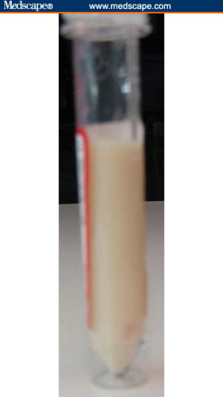
Laboratory data were remarkable for low hematocrit of 34%, low platelet count of 141,000, creatinine of 1.7, blood urea nitrogen of 55, mildly elevated liver enzymes (AST [aspartate aminotransferase] = 65, ALT [alanine aminotransferase] = 73, and alkaline phosphatase = 153), total protein of 5.9 and albumin of 2.9, and INR [international normalized ratio] of 1.2. The blood tests were negative for hepatitis A, B, and C. A CT [computed tomographic scan] of the abdomen and pelvis with oral contrast revealed massive ascites throughout the abdomen and pelvis, surgical clips, and a midmesenteric mass with calcification measuring 9 cm – which is larger than the prior examination size of 7.8 cm 14 months ago. Paracentesis was performed draining 4.5 L of milky white ascitic fluid on gross examination and had a triglyceride level of 1923 mg/dL on laboratory analysis (Figure). These results were consistent with chylous ascites. The chylous ascitic fluid was negative for acid-fast bacilli, had atypical or malignant cells, and had low amylase. These studies pointed toward sclerosing mesenteritis as the likely etiology.
Figure.

Test tube showing chylous (white) ascitic fluid.
Discussion: Sclerosing mesenteritis is a rare, idiopathic, and benign disorder of middle-aged or older adults (sixth to seventh decade of life), affecting the small bowel mesentery that is characterized by fat necrosis, fibrosis, and chronic inflammation.[1,2] The disease appears to be twice as common in men as in women.[3] The term sclerosing mesenteritis now represents an appropriate umbrella for numerous terms, such as mesenteric lipodystrophy, mesenteric sclerosis, retractile mesenteritis, and mesenteric panniculitis.[3,4] It poses a diagnostic challenge for physicians because it can be mistaken for malignancy,[5] particularly lymphoma, and is sometimes associated with diseases, including vasculitis, granulomatous diseases, retroperitoneal fibrosis, multiple myeloma,[6] fever of unknown origin,[7] pancreatitis, and carcinomatosis.[8,9] There have been reports associating sclerosing mesenteritis with idiopathic bile duct fibrosis simulating Klatskin's tumor[10] and occlusion of the inferior mesenteric vein.[11] Our case report has focused attention on a unique presentation in a patient with sclerosing mesenteritis. Chylous ascites is the accumulation of a milklike peritoneal fluid rich in triglycerides, due to the presence of thoracic or intestinal lymph in the abdominal cavity. It develops when there is disruption of the lymphatic system due to traumatic injury or obstruction (from benign or malignant causes). In Western countries, abdominal malignancy and cirrhosis account for over two thirds of all cases.[12,13] In contrast, infectious etiologies, such as tuberculosis, are responsible for the majority of cases in developing countries.[12,14] Other causes of chylous ascites include congenital, inflammatory, postoperative, traumatic, and miscellaneous disorders.[12] Congenital abnormalities of the lymphatic system and trauma should be considered as etiologic factors in children.[12] Diagnosis of chylous ascites can be readily made. The presence of a milky and creamy ascitic fluid with a triglyceride content above 200 mg/dL makes the diagnosis of chylous ascites.[12] In a recent article from the Mayo Clinic by Akram and colleagues,[3] 14% of sclerosing mesenteritis patients were found to have chylous ascites.[3] It is believed that direct mechanical compression by the mesenteric mass encasing the bowel, blood vessels, and lymphatics results in abdominal pain, bowel obstruction, ischemia, and chylous ascites.[3] Our patient had no history of cirrhosis, abdominal trauma, pancreatitis, and lymphoma, and the ascitic fluid was negative for acid-fast bacilli. There have been reports of treating chylous ascites with lowering the intake of triglycerides and substituting medium-chain fatty acids in the diet. There have been reports about the use of somatostatin and octreotide to treat chylous effusions in patients with yellow nail syndrome and lymphatic leakage caused by abdominal and thoracic surgery.[12,15,16] There have been no specific recommendations for treatment of chylous ascites in patients with sclerosing mesenteritis other than treatment of the primary cause. In our patient, dietary changes, colchicine, and steroids did not help during the follow-up period of 3 months, and the patient underwent palliative large-volume paracentesis for symptomatic improvement.
A paucity of cases of chylous ascites in sclerosing mesenteritis patients may hinder the study of different medications and approaches to treat this debilitating condition. Additional careful studies need to be performed to study the etiology of sclerosing mesenteritis to elicit the methods of treatment and avoid secondary manifestations of the disease itself.
Footnotes
Reader Comments on: A Clinical Case Study: Sclerosing Mesenteritis Presenting as Chylous Ascites See reader comments on this article and provide your own.
Readers are encouraged to respond to George Lundberg, MD, Editor in Chief of The Medscape Journal of Medicine, for the editor's eyes only or for possible publication as an actual Letter in the Medscape Journal via email: glundberg@medscape.net
Contributor Information
Manish Arora, Johns Hopkins University School of Medicine/Sinai Hospital Program in Internal Medicine, Baltimore, Maryland.
Ethan Dubin, Division of Gastroenterology, Sinai Hospital of Baltimore, Baltimore, Maryland.
References
- 1.Venkataramani A, Behling CA, Lyche KD. Sclerosing mesenteritis: an unusual cause of abdominal pain in an HIV-positive patient. Am J Gastroenterol. 1997;92:1059–1060. [PubMed] [Google Scholar]
- 2.Hirono S, Sakaguchi S, Iwakura S, Masaki K, Tsuhada K, Yamaue H. Idiopathic isolated omental panniculitis. J Clin Gastroenterol. 2005;39:79–80. [PubMed] [Google Scholar]
- 3.Akram S, Pardi DS, Schaffner JA, Smyrk TC. Sclerosing mesenteritis: clinical features, treatment, and outcome in ninety-two patients. Clin Gastroenterol Hepatol. 2007;5:589–596. doi: 10.1016/j.cgh.2007.02.032. [DOI] [PubMed] [Google Scholar]
- 4.Emory TS, Monihan JM, Carr NJ, et al. Sclerosing mesenteritis, mesenteric panniculitis and mesenteric lipodystrophy: a single entity? Am J Surg Pathol. 1997;21:392–398. doi: 10.1097/00000478-199704000-00004. [DOI] [PubMed] [Google Scholar]
- 5.McCrystal DJ, O'Loughlin BS, Samaratunga H. Mesenteric panniculitis: a mimic of malignancy. Aust N Z J Surg. 1998;68:237–239. doi: 10.1111/j.1445-2197.1998.tb04754.x. [DOI] [PubMed] [Google Scholar]
- 6.Goh J, Otridge B, Brady H, Breatnach E, Dervan P, MacMathuna P. Aggressive multiple myeloma presenting as mesenteric panniculitis. Am J Gastroenterol. 2001;96:238–241. doi: 10.1111/j.1572-0241.2001.03384.x. [DOI] [PubMed] [Google Scholar]
- 7.Sans M, Varas M, Anglada A, Esperanza Bachs M, Navarro S, Brugues J. Mesenteric panniculitis presenting as fever of unknown origin. Am J Gastroenterol. 1995;90:1159–1161. [PubMed] [Google Scholar]
- 8.Ege G, Akman H, Cakiroglu G. Mesenteric panniculitis associated with abdominal tuberculous lymphadenitis: a case report and review of the literature. Br J Radiol. 2002;75:378–380. doi: 10.1259/bjr.75.892.750378. [DOI] [PubMed] [Google Scholar]
- 9.Lim CS, Ranger GS, Tibrewal S, Jani B, Jeddy TA, Lafferty K. Sclerosing mesenteritis presenting with small bowel obstruction and subsequent retroperitoneal fibrosis. Eur J Gastroenterol Hepatol. 2006;18:1285–1287. doi: 10.1097/01.meg.0000243874.71702.21. [DOI] [PubMed] [Google Scholar]
- 10.Medina-Franco H, Listinsky C, Mel Wilcox C, Morgan D, Heslin MJ. Concomitant sclerosing mesenteritis and bile duct fibrosis simulating Klatskin's tumor. J Gastrointest Surg. 2001;5:658–660. doi: 10.1016/s1091-255x(01)80109-8. [DOI] [PubMed] [Google Scholar]
- 11.Seo M, Okada M, Okina S, Ohdera K, Nakashima R, Sakisaka S. Mesenteric panniculitis of the colon with obstruction of the inferior mesenteric vein: report of a case. Dis Colon Rectum. 2001;44:885–889. doi: 10.1007/BF02234714. [DOI] [PubMed] [Google Scholar]
- 12.Cardenas A, Chopra S. Chylous ascites. Am J Gastroenterol. 2002;97:1896–1900. doi: 10.1111/j.1572-0241.2002.05911.x. [DOI] [PubMed] [Google Scholar]
- 13.Rector WG., Jr Spontaneous chylous ascites of cirrhosis. J Clin Gastroenterol. 1984;6:369–372. [PubMed] [Google Scholar]
- 14.Jhittay P, Wolverson R, Wilson A. Acute chylous peritonitis with associated intestinal tuberculosis. J Pediatr Surg. 1986;21:75–76. doi: 10.1016/s0022-3468(86)80662-5. [DOI] [PubMed] [Google Scholar]
- 15.Widjaja A, Gratz KF, Ockenga J, et al. Octreotide for therapy of chylous ascites in yellow nail syndrome. Gastroenterology. 1999;116:1017–1018. doi: 10.1016/s0016-5085(99)70097-1. [DOI] [PubMed] [Google Scholar]
- 16.Shapiro AM, Bain VG, Sigalet DL, Kneteman NM. Rapid resolution of chylous ascites after liver transplantation using somatostatin analog and total parenteral nutrition. Transplantation. 1996;61:1410–1411. doi: 10.1097/00007890-199605150-00023. [DOI] [PubMed] [Google Scholar]


