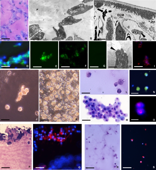
Figure 2. Analysis of the phenotype of cells recruited into the matrigel sponges by VEGF.
VEGF induces the mobilization of cells from tissues surrounding the MG implant into the biopolymer that was recovered and processed for structural, ultrastructural immunocytochemical analysis or seeded for cell culture. After 1 week in vivo the matrigel plug contained hematopoietic precursor cells stained with Giemsa solution (A) that were ultrastructurally similar (B) to those localized in capillary (c) and/or in the extracellular matrix (ECM) in stimulated leeches (arrowheads in C). These cells were small in size with large nucleus and scarce cytoplasm (B). Immunocytochemical characterization of cells infiltrating the MG supplemented with VEGF (D–G). The cells expressed several markers typical of hematopoietic/endothelial cells: CD34 (D), CD117 (E), CD31 (F); (G: negative control). The cells showed a migratory phenotype with degradation of ECM that was associated with their cytoplasmic projections (H, arrowhead), were also cathepsin B positive (I). After 1 week in vivo the MG infiltrated with precursor cells was removed and used to seed cell cultures (J–O). Phase-contrast (J, K) and May Grunwald Giemsa staining (L, M) micrographs, obtained 3 days (J, L) and 1 week (K, M) post-seeding, shows the increasing cell numbers from the initially few and scattered (J, L) to growth in clusters (K, M). Immunofluorescence detects expression of CD34 (N), a marker of precursor cells, of the cells label with BrdU indicating that these cells have cycled through S-phase (O). After incubation with Dil-Ac-LDL, those cells that localized in the superficial area of MG implant were Giemsa positive (P) took up fluorochrome-conjugated Ac-LDL (Q). Identical results were found within the monolayer of these cultured cells that were Giemsa and DIL positive (R, S). Taken together, these results strongly indicate that the cells migrating into the MG in vivo growing out in vitro consisted essentially of precursor/endothelial like cells. Bars: A 20 µm; B 2 µm; C 4 µm; D-N, P, Q 10 µm; O 5 µm; R, S 50 µm.

