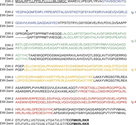
Figure 3. Alignment of EWI-2 sequence and the deduced EWI-2wint sequence.
Asterisk indicates the position at which an Arg (R) residue was introduced to make a furin cleavage site. Underlined amino acids correspond to the signal peptide of EWI-2. The four Ig domains are colored. Amino acid residues corresponding to the putative transmembrane domain are in italics and those corresponding to the cytoplasmic tail are bolded.

