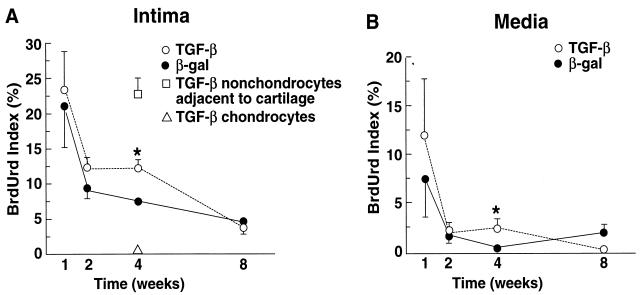
Figure 3.
Cell proliferation of TGF-β and β-gal arteries measured by incorporation of BrdUrd. (A) Proliferation in intima. At 4 weeks (but not at other time points), the difference in proliferative indices between TGF-β and β-gal arteries is significant (∗, P < 0.05). In the TGF-β arteries at 4 weeks, the proliferative index was elevated in nonchondrocytes adjacent to cartilage but was low in chondrocytes. (B) Proliferation in media. At 4 weeks (but not at other time points), proliferative indices in the TGF-β1 and β-gal arteries were significantly different (∗, P < 0.05). Data are mean ± SEM of three to five arteries/group.

