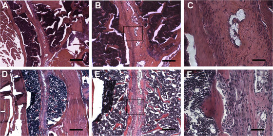
Figure 1.
Bilateral inflammation in sacroiliac joints of TnfΔARE mice. (A–C) Sacroiliac joint from a healthy C57BL/6 control. The lower two thirds of the joint consists of fibrocartilagenous tissue, and it is well defined and separated from the periost of the iliac bone. (D–F) Sacroiliac joint from a TnfΔARE/+ mouse on the C57BL/6 background. The presence of a mononuclear infiltrate within the sacroiliac joint with an increased number of blood vessels is shown. The fibrocartilagenous portion of the joint is invaded by mononuclear cells that protrude from the periost of the iliac bone into the BM. Paraffin sections are stained with hematoxylin and eosin. The boxes in B and E indicate the high-magnification areas shown in C and F, respectively. Bars: (A and D) 500 μm; (B and E) 200 μm; (C and F) 50 μm.

