Figure 2.
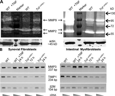
Mesenchymal cells from TnfΔARE mice are activated before disease onset. (A) Representative gelatin zymogram showing MMP9 and MMP2 secretion either from SFs (left) or IMFs (right) derived from WT and TnfΔARE/+ 4-wk-old mice. (B) Representative semiquantitative RT-PCR for MMP3 and TIMP1 expression in SFs (left) or IMFs (right) derived from WT and TnfΔARE/+ 4-wk-old mice. WT cells treated with TNF were used as a positive control in A and B.

