Figure 1.
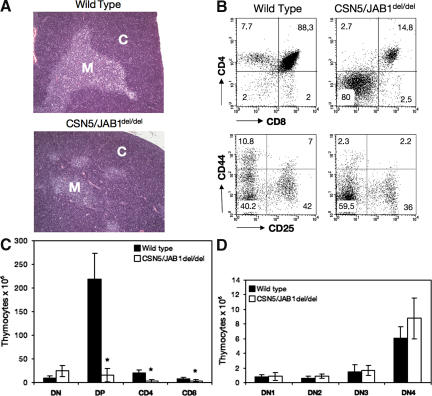
Impaired thymocyte development in CSN5/JAB1del/del mice. (A) Low magnification, hematoxylin and eosin–stained sections of thymi from WT (top) or CSN5/JAB1del/del (bottom) 5-wk-old mice show a greatly reduced medullary area, which is indicative of defective maturation of thymocytes in the absence of a functional CSN. Bar, 100 μm. M, medulla; C, cortex. (B) Thymocyte suspensions were stained with anti-CD4 plus anti-CD8 antibody to identify the major thymocyte subsets, and with anti-CD44 plus anti-CD25 antibody to detect the various subsets of DN thymocytes in WT (left) versus CSN5/JAB1del/del (right) mice. Percentage values for the various subsets are shown in the respective quadrants. (C) Thymic cellularity, as estimated by calculation of the number of thymocytes from individual thymi after electronic gating of the various subsets. Numbers are the mean ± the SD of values obtained from 25 mice per genotype at 5–6 wk of age. *, P < 0.001 (Student's two-tailed t test with equal variance). (D) DN subset distribution, calculated as in C, but after assessing the various DN subsets with anti-CD44 and -CD25 antibodies to identify DN1 (CD44+; CD25−), DN2 (CD44+; CD25+), DN3 (CD44−; CD25+), and DN4 (CD44−; CD25−) subsets.

