Figure 3.
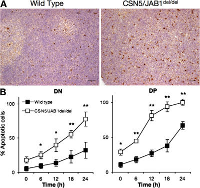
Increased apoptosis in developing CSN5/JAB1del/del thymocytes. (A) Immunohistochemical sections of 5-wk-old thymi from WT (left) or CSN5/JAB1del/del (right) mice stained with anti-cleaved caspase 3 antibody to detect apoptotic cells (as revealed by HRP immunoenzymatic reaction). Bar, 50 μm. (B) Isolated thymocytes from WT (filled squares) or CSN5/JAB1del/del (empty squares) mice were cultured in complete tissue culture medium for the indicated time points, after which the rate of apoptosis was assessed by flow cytometry as indicated in the Materials and methods section. Electronic gating was performed to quantify apoptosis in DN and DP thymocyte subsets. Data are the mean ± the SD of values obtained from eight independent experiments. *, P < 0.05; **, P < 0.001 (Student's two-tailed t test with equal variance).

