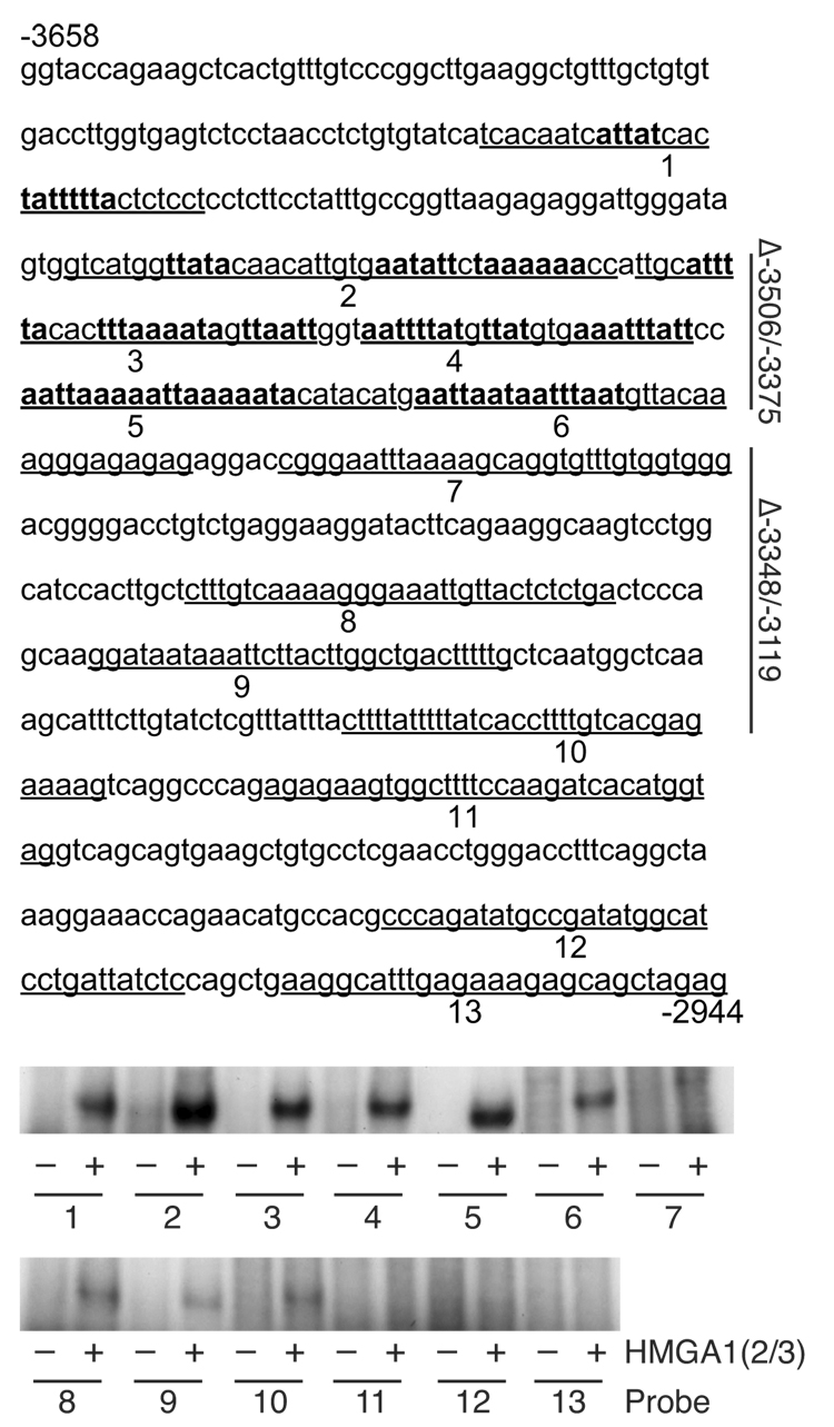
Fig. 4.
HMGA1 binds to the hNOS promoter. The hNOS2 5′-flanking sequence between −3658 and −2944 is depicted, with 13 DNA sequences (underlined and numbered) containing AT-rich regions used as probes for protein-DNA binding assays. The bottom panels show protein-DNA binding reactions using these probes after radiolabeling, in the presence (+) or absence (−) of synthetic HMGA1 peptide (HMGA1(2/3), 500 ng). Reaction mixtures of the probes and HMGA1(2/3) were then subjected to electrophoresis. Potential HMGA1 binding sites are denoted by bold type. A vertical line in the right margin designates regions −3506/−3375 and −3348/−3119, which are deleted (Δ) and tested functionally in Figure 5.

