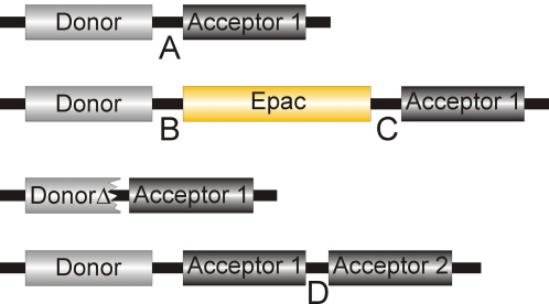
Figure 1. Schematic overview of the constructs used in this study.
Donor and acceptor fluorophore are connected by a peptide stretch (Linker A: SGLRSRYPFASEL) or by the Epac1(ΔDEP, CD) domain [1]. Within this stretch, the amino acids PF were replaced by the Epac domain itself, leaving linkers B: SGLRSRY and C: ASEL. For truncated donor constructs (CFPΔ and GFPΔ) GITLGMDELYK was deleted from the donor FPs and SGLRS from the linker. In tandem acceptor constructs the acceptors were separated by a supplementary linker (Linker D: PNFVFLIGAAGILFVSGEL) except for tdHcRed and tdTomato which have distinctive linkers, namely NG(GA)6PVAT) and (GHGTGSTGSGSSGTASSEDNNMA), respectively.

